


To date, science knows 280 types of worms that can develop and live in the human body and parasitize in various organs and tissues. The frequency of infection with human worms depends on the climatic and socio-economic conditions of certain areas (in underdeveloped countries, especially in tropical and subtropical areas, the extent of parasitic infections is much higher than in economically developed countries).
Ways of human infection with helminths
- Biohelminthiasis (infection by animals).
- Contagious helminthiases (transmitted from person to person).
- geohelminthiasis (diseases caused by parasites that carry out one of their life cycles on earth).
Factors that affect the manifestations of helminthiasis
- The way in which the parasite enters the body;
- The degree of adaptation of the helminth to the human body;
- population density (number) of parasitic individuals;
- The worm's habitat (tissue parasites live in the thickness of soft tissues, and luminal parasites live in the lumens of hollow organs). Some helminths in different phases have both lumen and tissue shapes. Larval and developmental stages of worms, as a rule, cause more severe pathological changes.
Without renewed infection, the number of adult parasites in the human body does not increase. This feature significantly differentiates helminthic invasions from diseases caused by bacteria, viruses, fungi, and protozoa.
Worms in humans: symptoms
Helminthiasis is a disease that is characterized by two stages of course (acute, from two weeks to two months) and chronic (from several months to several years).
Symptoms of the acute phase of helminthiasis
The first signs of the disease can appear at different times (mostly after 2-3 weeks, with ascariasis - after 2-3 days and with filariasis, the incubation period can last 6-18 months).
In the acute stage of parasitic invasion, the most characteristic symptom is an allergic reaction (antibodies are produced against antigens from migrating parasite larvae). People who are infected with worms often experience itchy rashes on the skin, which tend to recur, increase regional lymph nodes, generalized or local edema, muscle and joint pain can occur. In addition, migrating parasite larvae can cause chest pain, coughing, attacks of suffocation, upset stool, nausea, and vomiting.
At the same time, the acute phase of helminthiasis can be accompanied by more serious disorders (severe forms of pneumonia, hepatitis, allergic myocarditis, hepatosplenomegaly (enlarged liver and spleen), meningoencephalitis).
The number of eosinophils in the blood increases (eosinophilia) and the normal quantitative ratio between protein fractions is disturbed (dysproteinemia).
Signs of chronic helminthiasis
The symptoms of the chronic phase depend directly on which organ is "inhabited" by parasites, and their size and number play an important role.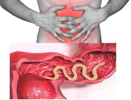 So if individual people parasitize in the intestine, the disease can be asymptomatic (with the exception of cases of infection with very large parasites). The characteristic signs of the chronic phase of intestinal helminthiasis are dyspeptic disorders. In children, the asthenoneurotic and pain syndrome are more pronounced. With a massive invasion of roundworms, the development of intestinal obstruction, obstructive jaundice and pancreatitis is possible.
Helminths consume all the substances necessary for their vital activity from the host's body and cause digestive disorders, impaired absorption of vitamins, minerals, carbohydrates, proteins and fats. At the same time, the waste products of worms inhibit the normal intestinal flora and reduce the body's immune system.
In people suffering from helminthiasis, the risk of malignant tumors increases significantly due to a weakened immune system and an increased cell division process (a result of the constant restoration of tissues damaged by parasites).
Types of helminths that parasitize in the human body
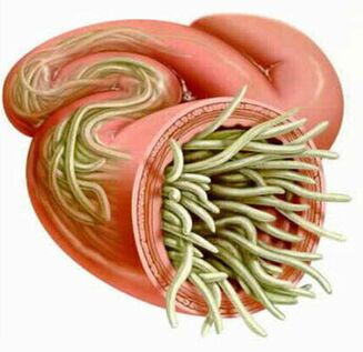
The causative agents of human helminthiasis are two types of worms: round (nematodes) and flat (tapeworms and flukes).
roundworms
pinworm
The parasites that cause enterobiasis are small (up to 10 mm) thin cavity worms with a gray-white color. Infection occurs through food (through the mouth). The reason for this is dirty hands. The eggs of the parasite can be in the ground, on the wool of infected animals, unwashed vegetables and fruits, etc. At the same time, with enterobiasis, there are often cases of self-infection (especially in children), due to scratching the itchy areas and the subsequent ingestion ofEggs.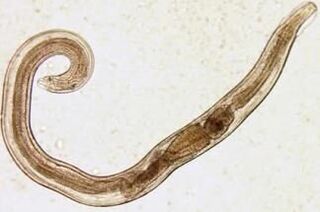 The pinworm larva develops in the digestive tract within two weeks. After the worm grows up, it parasitizes in the lower sections of the small and upper parts of the large intestine.
Even in the larval stage, the pinworm begins to damage the body of its host and to produce enzymes that irritate the intestinal walls and lead to the development of an inflammatory process. Adult parasites adhere to or penetrate the deeper layers of the intestinal lining, disrupting its integrity and contributing to the attachment of a secondary bacterial infection. With pinworms perforating the wall of the small intestine, peritonitis can develop. Due to the irritation of the intestinal receptors, the motor and secretory functions of the gastrointestinal tract are also disturbed, which leads to the formation of gastroduodenitis, enteritis, etc. In childhood, long-term enterobiasis can lead to nerve disorders and a delay in physical development.
Ascaris
Ascaris is a large spindle-shaped parasite of red-yellow color that reaches 40 cm (women) and 15-25 cm (men) in adulthood. Without suction cups or other fastening devices, the roundworm can move independently towards the food masses. The eggs laid by the female of the parasite are excreted with the faeces.
Infection with ascariasis occurs when ripe eggs are swallowed with water or unwashed vegetables and fruits with soil particles. After the eggs enter the intestine, they develop into mature larvae. Then they penetrate the intestinal wall, reach the heart through the bloodstream, and from there go to the lungs. The roundworm larvae return to the oral cavity through the respiratory tract through the pulmonary alveoli. After repeated swallowing, the parasite reaches the small intestine, where it develops into an adult. The worm lives 12 months, then dies and is excreted with the feces. Both one and several hundred individuals can live in the intestine of a host.
In the intestinal phase of their existence, roundworms, which have the ability to spiral movements, can penetrate even the narrowest openings. This feature of the parasite often leads to the development of rather serious complications (obstructive jaundice or pancreatitis). Allergens released by roundworms can cause severe allergic reactions. Large numbers of adults can cause intestinal obstruction, and worms getting into the airways sometimes cause choking hazards.
Vlasoglav
Vlasoglav, the causative agent of trichocephalosis, is a white helminth that parasitizes in the initial section of the large intestine and reaches a size of 4 to 5 cm. The parasite feeds on blood and tissues of the rectal mucosa.
The whipworm eggs placed on the intestinal walls by the female come out together with the feces. Their development takes place in the environment (optimally in the soil). Eggs with the larvae of the parasite that have matured in them get into the body through food, dirty hands, with water or unwashed vegetables and fruits.
With a small number of worms, trichocephalosis is asymptomatic. At a severe stage (with massive invasion), the patient develops abdominal pain, severe diarrhea develops, sometimes accompanied by rectal prolapse. This condition is most often seen in debilitated children. With a moderate phase of trichocephalosis, growth retardation of a child is possible.
Trichina
The causative agent of trichinosis is a small round helminth with a length of 2-5 mm. Infection occurs when poorly roasted meat (pork, bear meat, wild boar) is eaten.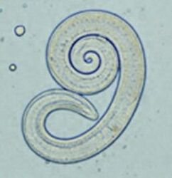 The larva of the parasite penetrates the intestine and matures in 3-4 days to the state of a sexually mature individual. The lifespan of the worm is 40 days, after which the parasite dies. By piercing the intestinal wall, the larvae enter the bloodstream and are transported to all organs in the human body, settling in the muscles. In this case, the muscles of the respiratory and facial muscles, as well as the flexors of the limbs, are most often affected.
In the first few days after the invasion, patients complain of abdominal pain. Then, after about 2 weeks, the body temperature rises to 39-40 ° C, itchy rashes appear on the skin, muscle pain develops, and the face swells. During this time, there is a significant risk of death if there is a massive infection. After about a month, the patient will recover. The parasite is encapsulated in a spiral and dies within two years.
Hookworm and Nekator
These two parasites are similar in their biological properties as well as in the diseases they cause. In this regard, it is common to combine them under a common name (hookworm). Worms that reach 10-15 mm in length parasitize in 12-p. Intestines. It should be noted that this is one of the most common, but at the same time quite rarely discovered parasites. Worm larvae enter the human body through the skin through contact with contaminated soil. When they enter the bloodstream, they travel like roundworms to the lungs and then along with the sputum through the bronchi to the digestive tract. The ankylostoma parasitizes in the intestine and attaches itself to the intestinal wall. The parasite, which feeds exclusively on blood, bites through the blood vessels that penetrate the mucous membrane and injects an anticoagulant component there. On average, an adult can ingest 0. 05 to 0. 35 ml of blood per day. Therefore, the most characteristic symptom of this helminthiasis is iron deficiency anemia, as well as a change in the ratio of protein fractions (dysproteinemia).
flatworms
Wide ribbon
This is one of the largest helminths with a length of 10-20 meters. The disease caused by this parasite is called diphyllobothriasis. The worm's development cycle begins with freshwater fish or crustaceans. The larva enters the human body along with eggs or infected fish fillets, which is the ultimate owner of the broad tapeworm. The parasite reaches the small intestine, clings to its wall, and grows into a mature individual within 20 to 25 days.
Diphyllobothriasis occurs against the background of digestive tract disorders and B12 deficiency anemia.
Liver fluke
The parasite that causes opisthorchiasis is a flat worm that reaches 7-20 mm in length. It should be noted that more than 50% of cases of liver fluke (also called cat fluke) infections occur among residents of Russia. The larvae of the parasite begin to develop after the eggs enter fresh water (from the snails that ingested them). Then they penetrate the body of fish (carp, crucian carp, bream, roach). Human infection occurs when contaminated fish meat is eaten that has not undergone adequate heat treatment. The larva of the liver fluke from the small intestine penetrates the biliary tract and the gallbladder and fixes itself there with the help of two suction cups.
In the acute phase of helminthiasis, the patient has pain in the upper abdomen, the body temperature rises, nausea develops, muscle pain, diarrhea and rashes are possible. The chronic course of opisthorchiasis manifests itself in symptoms of hepatitis, inflammation of the biliary tract, cholecystitis, disorders of the digestive tract, nerve disorders, weakness and increased fatigue. The parasite leads to the development of irreversible changes, and even after its expulsion, the patient is not subject to chronic inflammatory processes and dysfunction.
Beef and pork tapeworm
These almost identical parasites reach a length of 5-6 meters. Infection with teniarin pants and teniasis occurs from eating cattle or pork that has been infected by the Finns (one of the intermediate forms of helminthiasis). Viable fins, presented in the form of whitish bubbles 0. 5 cm in size, attach to the wall of the human small intestine and grow up in 3 months. The ribbon parasite, which consists of more than 2000 segments, is constantly growing. In this case, the end segments containing eggs break off and move independently along the colon to the anus, and then crawl out of the anus or are released into the external environment along with the feces. The most characteristic symptoms of helminthiasis are digestive tract disorders.
Echinococcus
For this parasite, a person is an intermediate host. The worm parasitizes the human body in the form of fins. The last owner of Echinococcus is a wolf, dog or cat.  The infection occurs due to diet through contact with animals and with environmental objects that are sown with Echinococcus eggs. After entering the intestine, oncospheres (larvae with six hooks) develop from them. They enter the bloodstream from the intestine and are transported through the body.
The worm's "preferred" parasitic sites are the liver and lungs. By settling in these organs, the larva turns into a finn (echinococcal cyst), which, as it grows, begins to destroy nearby tissues. Often in the diagnostic process, echinococcosis is confused with a tumor of benign or malignant origin. In addition to mechanical shocks (squeezing organs and blood vessels), an echinococcal cyst rupture sometimes occurs. This condition can cause toxic shock or the formation of several new cysts.
Alveococcus
This parasite, considered a type of echinococcus, is the cause of one of the most dangerous helminthiasis (alveococcosis), the severity of which is similar to cirrhosis and liver cancer. Infection occurs when oncospheres (eggs with mature larvae) enter the intestines. There the embryo leaves the egg and penetrates the intestinal walls and enters the bloodstream. In addition, with the blood supply, the parasite spreads to all tissues and organs of the body (most often it is localized in the liver). This is where the main development stage in the larvae begins (a multi-chambered bladder, laurocyst is formed). Each chamber contains the parasite's embryonic head, which gradually evolves. Laurocysts are very aggressive formations that constantly grow due to the enlarging bladders and, like cancer metastases, can also grow into the liver. Necrotic changes due to dysfunction of blood vessels are subject to necrotic changes in nearby tissues. The alveococcus spreads to nearby structures and forms fibrous nodes with inclusions of multi-chambered bubbles. This condition can persist for several years, so it requires mandatory surgical intervention.
Diagnosis of helminthiasis
Diagnosing helminth invasions involves the following activities:
- a thorough medical history that will help to find out the possible causes of infection;
- Laboratory tests of feces, blood, intestinal contents 12p, rectal and perianal mucus, muscle tissue, lung sputum, bile. The analysis can reveal eggs, segments or fragments of parasites. At the same time, an increased level of eosinophils in the blood is also a signal of the presence of helminthiasis.
- When diagnosing diseases caused by larval stages or tissue parasites, serological studies are carried out (ELISA, RSK, indirect agglutination reaction, immunofluorescence analysis, etc. ).
- Ultrasound, CT, and endoscopic exams are prescribed to detect helminths that affect liver tissue.
Human worms: treatment
In the acute phase of a parasitic infection, the patient is prescribed detoxification and desensitization therapy. In severe cases of illness (trematodes of the liver, trichinosis), glucocorticoids are used according to medical indications.
As a drug of a specific therapy, special anthelmintic chemotherapeutic agents are prescribed, taking into account the type of pathogen.
In parallel, the patient is recommended to take antihistamines and enterosorbents. The final stage of treatment includes the use of probiotics, which normalize the intestinal flora.
A special, economical diet is also prescribed (food should be digestible and low in fat).
During the antihelminthic therapy, the patient must pay close attention to personal hygiene (to avoid re-infection). At the same time, with many helminthiasis, all family members and people who are in constant contact with the infected must be treated.
Prevention of helminthiasis
- Maintaining personal and public hygiene;
- Strict adherence to cooking technology;
- Regular examination and preventive treatment of pets;
- Thoroughly washing fresh vegetables, fruits and herbs;
- Correct handling of river fish;
- Avoid eating raw, lightly salted and dried fish.